U.S. flag

An official website of the United States government

Official websites use .gov
A .gov website belongs to an official government organization in the United States.

Secure .gov websites use HTTPS
A lock ( ) or https:// means you’ve safely connected to the .gov website. Share sensitive information only on official, secure websites.

Case Study: Jeff

A button to download a simplified PDF version of this case

Welcome to Your Patient!

Jeff's photoJeff, 46 year old American male of Scots descent has come to you for a second opinion...

Previous Diagnosis: Cirrhosis of the liver and Type 1 Diabetes, due to chronic alcoholism

Key Symptoms: Elevated liver enzymes, excess ascites fluid and an enlarged spleen (splenomegaly)

In the two months since his initial diagnosis, the prescribed insulin treatment and significant dietary changes have not seemed to improve things much. Indeed, his condition has deteriorated including severe fatigue and some vision issues. He claims he is not, nor has he been an alcoholic – used to have 1-2 glasses of beer a few times a week and had not had anything for a while before being assessed – so he really doesn’t think this diagnosis makes any sense.

Subsequently, Jeff discussed his case this a friend who is a retired pathologist who suggested that he get checked for a genetic condition: Hemochromatosis.


Researching the Referral

In this scenario, Jeff was referred to the genetics clinic because the suggested diagnosis suggested that he might have a potentially serious genetic condition. If a known pathogenic genetic variant is found, it can validate the diagnosis and provide additional patient-specific information that might help customize his case management plan.
Jeff's Referral icon

To learn more about a case, please click on the Referral icon to open the form. 

Guidance Form icon
In the Patient Case Review guide sheet provided by your attending:  Notate some key clinical features listed for Jeff as well as a preliminary diagnosis.

This is the beginning of preparing for a possible molecular case presentation.


To learn more about a possible genetic disorder that is consistent with what Jeff is experiencing, search in NCBI's MedGen Database with the several of Jeff's clinical features:

cirrhosis AND diabetes AND ascites AND splenomegaly


Webpage iconIf you need it, you can click here to get to a direct link for the Medgen record page.

Try this!

 
Make an observation iconBased on the results of the search, what does it suggest is a possible diagnosis for Jeff? How does this correspond to the preliminary diagnosis listed in the referral?


The "Disease Characteristics" section of this record displays a summary from a specific GeneReviews® Chapter on the NCBI Bookshelf.

Make an observation icon
Scan the summary
and consider accessing and reading the entire GeneReviews® Chapter! 


A helpful webpage -icon.Note:  GeneReviews® is a project run by the University of Washington producing expert-authored, point-of-care information with clinically relevant and medically actionable information for inherited conditions.  It is an incredible review article-type of resource, and is thus featured in it's own section on relevant MedGen records - as an abstract with links to key sections.


Interpretation icon
Based on what you know about Jeff and what you've read, what do you think is a reasonable preliminary diagnosis?  (Yes, you'd probably want to do some more research on this and this MedGen record has links to a lot of helpful information in PubMed and elsewhere...but for this particular excercise, go with your "gut" instinct.)

Guidance Form icon
In the Patient Case Review guide sheet:  Notate your preliminary diagnosis.


Under the GeneReviews® Summary, click on the heading "Diagnosis" read through that section through the "Establishing the Diagnosis" sub-section.

Make an observation icon
What other disorders might you consider in your differential assessment?


Another good source for information about related disorders is in the “Term Hierarchy” section of this record. Here you can see a few sub-types of Hemochromatosis. Click the names of the disorders to open the MedGen records to read about each sub-type.  Note at the top is a gene or genes that have been associated with the disorder.

Make an observation icon
Find the gene or genes that is listed as being associated with each subtype.


Interpretation iconWhat do you think is one key parameter that you can use to differentiate the sub-types of Hemochromatosis?  (Hint:  This is a workshop about the impact of genetic variants on gene products.)


Picture of jeffYou discuss the possible diagnosis with Jeff.  He has heard that this is may be a genetic condition. He has a twin sister and a brother and is concerned that they may be affected. He’d like to have a DNA test to see if the cause of his disorder is genetic and also see if he can find out specifically what is wrong with him on a personal level, even though it may not change his prognosis or therapy.

You decide to move forward with ordering a genetic test and send an order to the Genetic Testing Laboratory.


What does the patient's genetic test report say?

Jeff went to the laboratory, provided a sample which was analyzed and the results have been sent to you for consultation. To see Jeff's genetic test results, please click on the Test Results.
Jeff's Genetic Test icon
Read it over and fill in below what you can glean from the report with regard to any genetic variants found and anything they laboratory might be asserting about how this relates to the preliminary diagnosis.

Guidance Form icon
In the Patient Case Review guide sheet:  Notate specific genetic variations that have been identified as well as what the laboratory asserts about their classification (i.e. Pathogenic, Likely Pathogenic, Likely Benign, Benign, or Variant of Uncertain Significance).


A helpful webpage -icon.Note:  A great central resource to find genetic tests for a condition-of-interest, is the NIH Genetic Testing Registry. You can search it directly from the resource's homepage or you can link to relevant genetic tests directly from a link on the right-hand side of a MedGen disorder page.


What is currently known about the identified variant?

Genetic testing laboratories attempt to stay up to date with what is known about the genetic variants that they are assessing. However, it is sometimes valuable to quickly consult with national database of clinical variants (NCBI's ClinVar database) to learn what other organizations have asserted/interpreted for that variant, if anything. In addition to information from testing laboratories, ClinVar receives curated interpretations from authoritative sources such as ClinGen, ACMG and disorder-specific specialist panels.

To learn more about the genetic variant identified in Jeff, search NCBI's ClinVar database with:
HFE Cys282Tyr

Webpage iconIf you need it, you can click here to get to a direct link for the ClinVar record page.                     Try this!

Look over the ClinVar records to see information submitted to NCBI's ClinVar from clinical laboratories, expert panels and clinical genetics organizations. 

Make an observation iconWhat is the listed Clinical Interpretation information for this variant?  

Click on the Conditions tab to learn more about specific disorders and the interpretations as they relate to each entry type.


Interpretation icon
Based on the submitted data, what do you think the most likely interpretation classification is for the disorder you are assessing? How does this relate to what the Genetic Testing Laboratory's Report asserted?

 
Guidance Form iconIn the Patient Case Review guide sheet:  Notate the asserted interpretation classification as listed in ClinVar.
 

Look in the Variant Details tab 
to find biology-related information about this variant.
Make an observation icon
To help map the genetic variant in the last section of this exercise, find the HGVS notations which precisely define the variant's position in different biological molecules.


Guidance Form iconIn the Patient Case Review guide sheet:  Notate the HGVS term for it's position in the gene (NG_###### g._______) and in the first protein information listed (NP_###### p.________) representing the MANE version of the protein encoded by the gene. 


What is currently known about the identified gene?

If a particular gene has been implicated in a genetic test results indicating a pathogenic variant exists in a patient, it is often helpful to understand what that gene is, what its normal function is, where it is found (cellular and tissue expression patterns), and other sources of accessible information, such as links to relevant scientific literature.

NCBI's Gene database aggregates data from many NCBI databases as well as other high-quality resources to provide information and links to help users find and understand what is currently known about a particular organism's gene.

To learn more about Jeff's impacted gene, search NCBI's Gene database with the gene symbol indicated on the genetic test result and find the record for the human version.

Look over the information about the gene that was implicated in the genetic test result. In the Summary section, read the Summary provided by the NCBI RefSeq project.

Look for the data icon
What is the normal function for the gene product? What else has been noted? 


Guidance Form icon

In the Patient Case Review guide sheet
:  Notate what you feel is important to know about the normal version of this gene product.


Navigate to the Expression Section of this record.

Look for the data iconExamine the histogram of expression levels for specific tissues. Note there are several tissue sets from different experimental studies available to view, by clicking and selecting to display the results in the pull-down menu.

Interpretation icon

In which tissues is this gene normally expressed?


Guidance Form icon

In the Patient Case Review guide sheet
:  Notate the tissues in which you would expect this gene to play a physiological role.


In the Gene Ontology Section of this record is a set of annotations for where this gene product is likely to be found within a cell (Component), what processes it is often involved in (Process), and what it does (Function).
 
Make an observation iconWhat type(s) of process(es) is/are this protein normally involved with?
What specific function(s) does this protein have?
In which component(s) (sub-cellular location) is this protein normally found?


Interpretation icon
Does this make sense based on the Gene Summary of the Gene that you found above? 


A helpful webpage -icon.
Note:  The Gene Ontology project, a.k.a. GO, is coordinated by an international consortium. It is an incredible resource with a very helpful standardized set of terms which is helpful to understand genes and also for computational research! 


Guidance Form iconIn the Patient Case Review guide sheet:  Notate GO annotations that correlate with what you've learned about the normal function(s) and locations of this gene product. Indicate any that seem to contradict or are new pieces of information that might be helpful.


Let's put this all of this together....
Interpretation icon

Does what you've found above make sense based on the patient's symptoms and personal history? 


A helpful webpage -icon.Note:  On the NCBI Gene page, there are links in Bibliography and GeneRIF Sections to relevant PubMed records so you can learn even more.....   


Click here to see some additional helpful information I've pulled it together from an old molecular biology textbook available on the NCBI Bookshelf!
  Iron transport and the Transferrin Cycle:
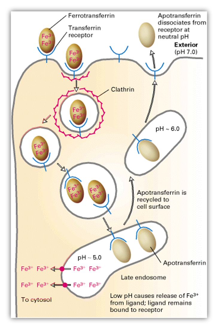
  • Figure showing "The Transferrin Cycle" from the textbook "Molecular Biology of the Cell" on NCBI's Bookshelf.Iron is an essential nutrient required for the synthesis of hemoglobin, cytochromes and many other proteins.
  • Iron is transported around the body by the protein Transferrin (often called Apo-Transferrin when not carrying Iron ions and Ferro-Transferrin when complexed).
  • Ferro-Transferrin binds to target cells via the Transferrin Receptor causing internalization and cellular uptake of the complex.
  • A localized pH change frees the Iron ions which are then released internally, while the Tranferrin (or Apo-Transferrin) is returned to the outside of the cell to repeat it's role in Iron transport.
  • To prevent possibly damaging high levels of intracellular Iron ions, the HFE Protein regulates the cellular uptake of the Ferro-Transferrin complex.


Map the variant through the bioinformatic flow!

Now that we understand which gene may be affected by the presence of the detected variant, mapping the variant through the central dogma of molecular biology can help indicate at which point it has its strongest impact.

Note: Currently, genetic testing laboratories most often use genomic DNA for their genetic tests - identifying a variant by its position on a particular chromosome. They most often report variants found in gene regions, since currently this is where most research has been focused on to explain impacts on human biology.

Observe iconClick here to review an overview of the central dogma and genetic variation. Graphic showing the bioinformatic flow steps for variant impact
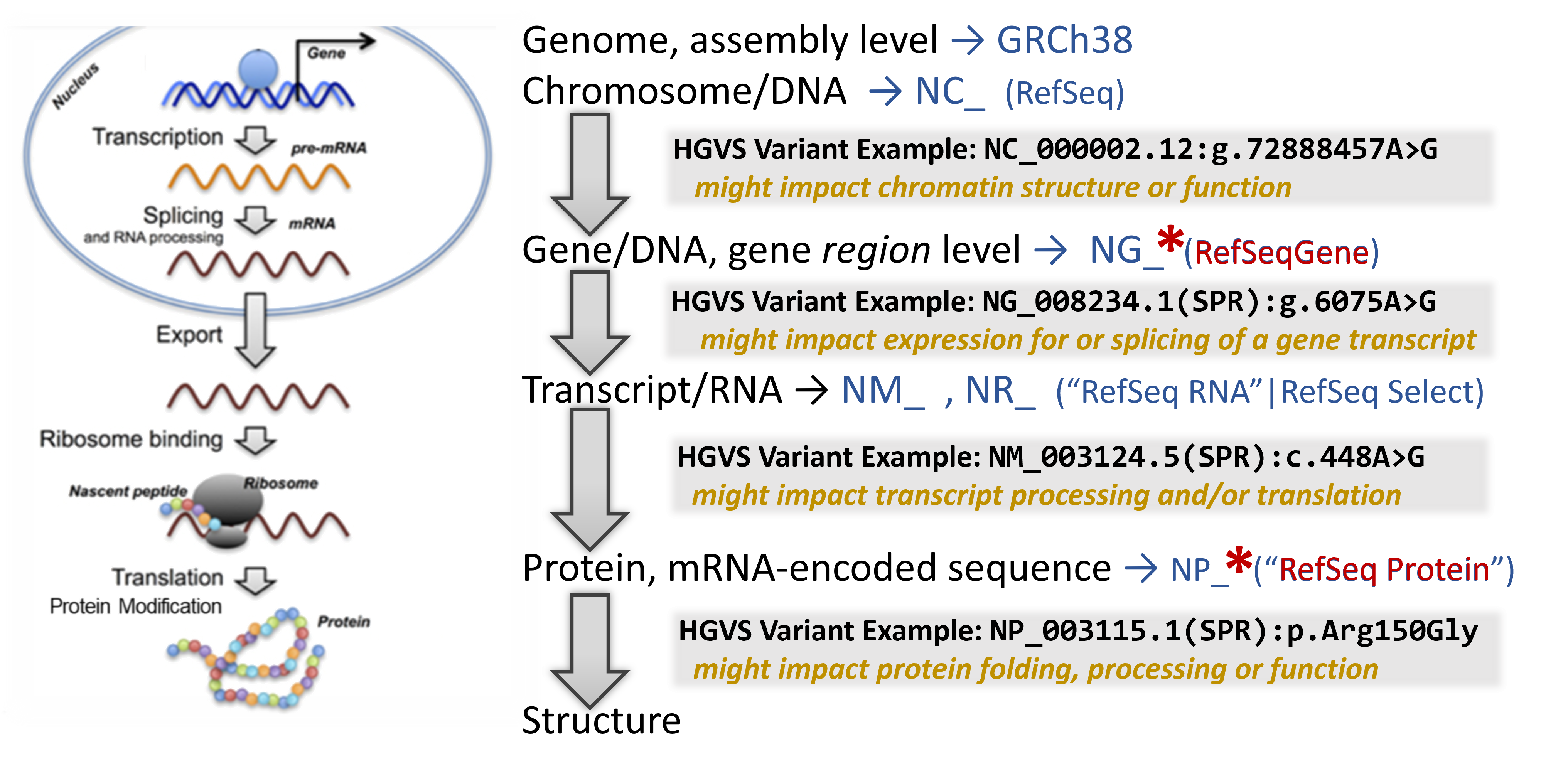
In addition to a lot of helpful aggregated information, NCBI's Gene database provides links to visualization tools which can help to identify where a variant is located in several critical biomolecules. 

To learn about the molecular impact of the genetic variant, begin your search on the relevant NCBI Gene record.  Then sequentially click on several helpful linked resources on this page to map the location and infer it's impact through this bioinformation flow
 
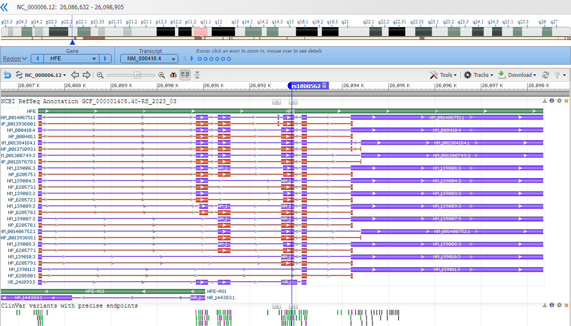
Genome Data Viewer shows the chromosome zoomed into the gene region. Click on the link in the right-hand side of the page to see it.

Observe iconClick here for a view of the GDV display and a link. Image of GDV view - HFE with variant marker
 

Note that you are looking at a portion of the chromosome (the accession shown is an NC_).
Make an observation icon
Which chromosome do you think you are looking at? i.e. which one is this gene encoded on?

Zoom into the location of the variants by entering an HGVS term, such as: NG_008720.2:g.10633G>A into the top-left text box to search for the variant location.

Where is the variant located in relation to the indicated gene?  (near a gene? upstream or downstream from the gene? in the gene region?)

Make an interpretation iconBased on the location of the variant and the type - what impact do you think it might have on the chromosome? What about the gene or genes in that region?


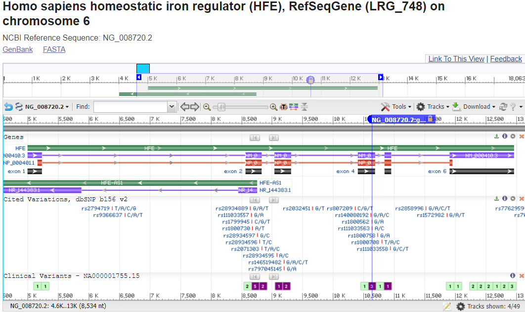
RefSeqGene has a Graphics view that shows the gene region, including some areas upstream and downstream. Click on the RefseqGene link in the right-hand side of the page, and then - under the title of the record - click on Graphics.

Observe iconClick here for a view of the RefSeqGene Graphic display. Image of RefSeqGene view - HFE gene with variant marker


Make an observation iconNote that you are looking at a small region of the chromosome focused specifically on the gene with a bit upstream and a bit downstream. (the accession shown is an NG_).

To zoom into the location of the variant, you can put that HGVS term NG_008720.2:g.10633G>A into the top-left text box to search for the variant location.

Where is the variant located in relation to the gene structure?
 
(in the upstream or downstream region surrounding the gene? in the 5'-untranslated portion of the transcript portion? near a splice site? in a coding exon?)

Make an interpretation iconBased on the location of the variant and the type - what impact do you think it might have on the gene expression and resulting transcript?


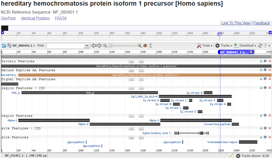
RefSeq Protein Graphic view shows the protein sequence, including conserved domains and other regions.   
Make an observation iconClick on the Refseq Protein link in the right-hand side of the page, and then look for the record labeled  hereditary hemochromatosis protein isoform 1 precursor [Homo sapiens] with the accession that matches what you found in ClinVar: NP_000401.1. (Hint: Likely the last one!)  And fnally, click Graphics to get to the interactive graphic view.

Observe iconClick here for a view of the RefSeq Protein Graphic display. Image of RefSeq Protein view - HFE with a variant marker

Note that you are looking at the full length of the protein sequence itself in this view. (the accession shown is an NP_).

To zoom into the location of the variants, you can click on the magnifying glass in the upper-left above the graphics display and enter HGVS terms such as: NP_000401.1:p.Cys282Tyr in the top-left text box to search for the variant location.
In this RefSeq Protein Graphics view, you can mouse-over some lines, such as region & site reatures - CDD to read informational pop-ups about what you are seeing.  You can even click on them to learn more about the functional regions.

Additionally, click on "Identify Conserved Domains"in the upper-right to learn more also and even see a graphic that more clearly the exact residues that play a specific role in this protein (although you'll have to use the protein sequence ruler to figure out where variants are in this view).

Observe iconClick here for a view of the Conserved Domains display.
Image of Conserved Domains view of the SPR protein
 
Make an interpretation iconWhere is the variant located in relation to the protein sequence and it's annotated functional domains? 


For many gene products such as this one, you can even see the 3D Structure of the protein, sometimes substrates and ligands and sometimes full complexes and map the location of the genetic variant impact within it.
 
Observe iconClick here to see something pretty cool! Image of the 3D structure of HFE with key variants highlighted The 3D crystal structure for the HFE Protein
(PDB accession: 1DE4) in NCBI’s Cn3D Viewer.
The Cys282 position is shown in a yellow and participates
in a disulfide bond (with another cysteine side chain,
orange stick) holding together two beta sheets in an Ig
Fold.

What would the change of that cysteine do to the structure?
What might be the impact on the ability of this protein to function?

HINT:  De Almeida, SF, and M De Sousa. “The Unfolded Protein Response Haemochromatosis.” Journal of Cellular and Molecular Medicine. PMC. Web. 7 Sept. 2016.

To understand the role of the HFE protein in biology, let’s look at two other 3D Structures that arerelated to this protein’s function and purpose (PDB accessions 1SUV – Transferrin Receptor complexed with Ferro-Transferrin & 1DE4 – Transferrin Receptor complexed with the HFE protein).
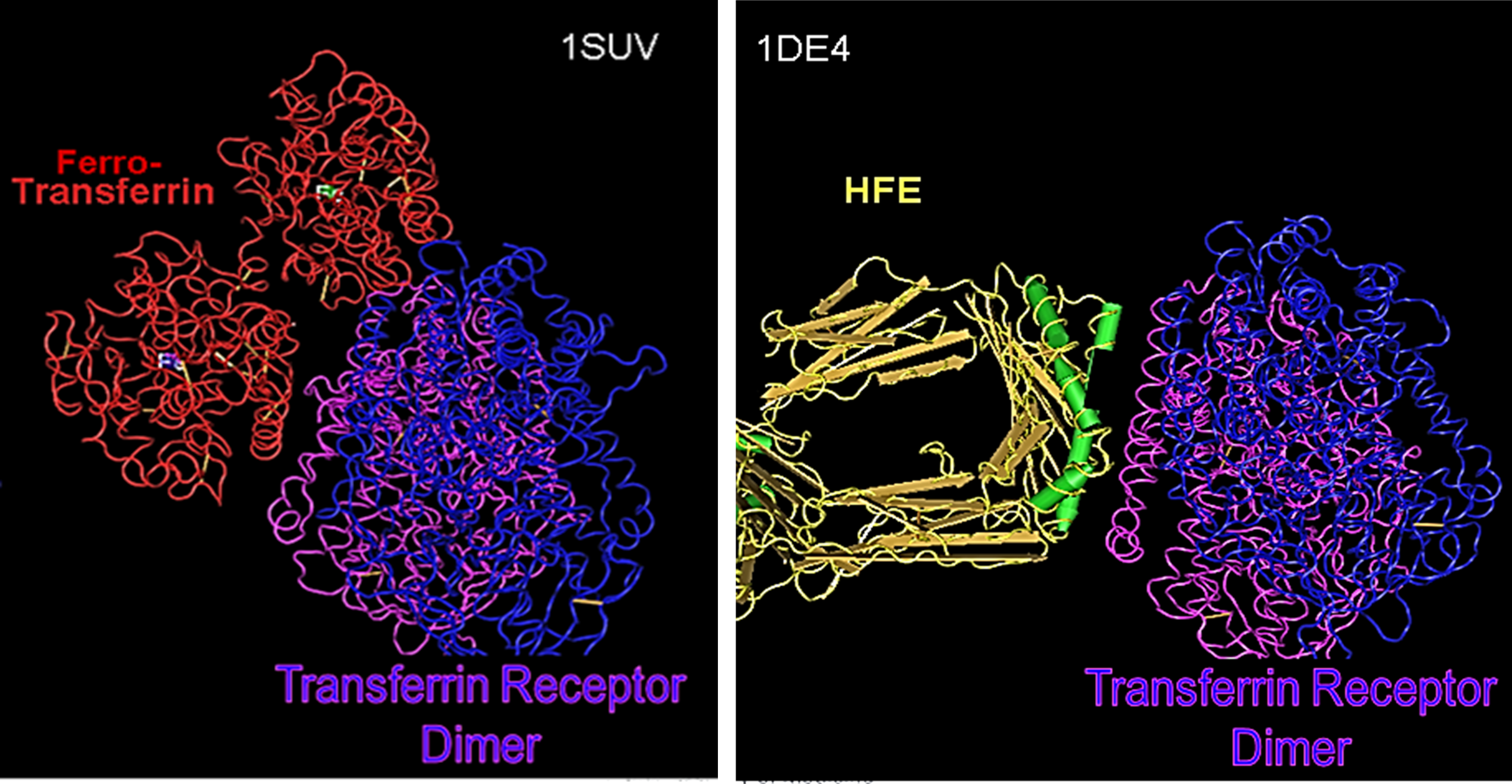
Image of the 3D structure of HFE with key variants highlighted

In both structures, the Transferrin Receptor is shown in purple & blue. On the left in red, you see how it complexes with Transferrin (ferro-transferrin means it is carrying its Iron ion payload).  On the right in yellow, orange & green (secondary structures – which are also shown in the picture of #9), you see how the Transferrin Receptor complexes with a homodimer of the HFE protein.

How do you think HFE regulates the interaction of the Transferrin Receptor with Ferro-Transferrin? 
What would happen to this if the HFE protein is encoded with the p.Cys282Tyr variant?


Make an interpretation iconBased on the location of the variant and the type - what impact do you think it might have on the gene's ultimate product - the protein?


Guidance Form iconIn the Patient Case Review guide sheet:  Identify the ultimate biological molecule where the genetic variant is found (chromosomal region, gene region, gene, transcript, protein or protein structure), and describe how it may impact the structure and/or function of that molecule.


Let's put it all together to understand what is happening in the patient!

Jeff's photoJeff would like to have some answers.

Click here to review some things you may want to consider when formulating the answer to his questions.
          • Which gene is impacted by the genetic variation and what does the gene product normally "do"?
            • what is it's biomolecular function?
            • what is it's impact on cellular physiology? 
            • in which cells/tissues is the gene product usually expressed?
          • Based on the patient's variation(s):
            • what do you think this would do to the gene product's structure and biomolecular function?
            • what would this do to cellular physiology?
            • what tissues or organs impact be impacted?
          • Based on the proposed impacted-tissues/organs, may some of the the patient's symptoms be explained by this?  (validating her experience)


Summary icon
What do you think is happening in your patient?

  Notes
Diagnosis  
Genetic Variation(s)  
Proposed Molecular Mechanism of Variant Impact  
How does this relate to the phenotype?  


Take-away message!


Workflow:   We've practiced this same step-by-step process to learn more about a different patient's genetic variant.

Genetic disorder & molecular pathology:  Missense variants involving cysteine residues, in particular, can have a devastating effect due to their key role in forming structure-stabilizing, covalent disulfide bonds across spans of a protein sequence. The loss of this structural cross-beam support can cause the protein to unfold, often disrupting at least part of the protein's ability to function and sometimes triggering an endogenous "unfolded protein response" which activates a process to destroy the unnaturally unfolded protein.

In this case, the HFE protein is destroyed and with it, it's ability to bind to and inhibit the Transferrin Receptor's import of Ferro-transferrin. Thus, the cellular import continues unregulated and the cell accumulates dramatically increasing levels of highly reactive iron ions. Over time, these cause damage to the cell and if it is not reversed and/or if the cell cannot undergo apoptosis (a clean, programmed cell death), then significant DNA and protein damage can accumulate and induce the cell to undergo neoplastic transformation. The most notorious cancer eventually developed by those with Hemochromatosis, being hepatocellular carcinoma.


ANSWER
Coming soon icon

Last Reviewed: August 22, 2023