

Step 6: Exploring Protein Sequence and Structure Data
Task(s): Visually identify sequence variation between IMA1 proteins in our three yeast species, and find the 3D structure of the IMA1 protein for further examination.
Background on NCBI Resources Used:
NCBI BLAST graphical results options: The web BLAST interface provides many options for visualizing and summarizing the results of a search. Of these, we are going to make the most use of the Alignment and MSA (Multiple Sequence Alignment) viewer options. More about the MSA Viewer, which can be used on its own, can be found starting from the NCBI MSA Viewer landing page. You will use these tools to identify variation between several IMA1 sequences.
Structure Database: NCBI’s Structure Database stores three dimensional macromolecular structure information. Three dimensional structures provide a wealth of information on the biological function and the evolutionary history of macromolecules. They can be used to examine sequence-structure-function relationships, interactions, active sites, and more. We are going to find and examine a Structure database record for an IMA1 protein.
Macromolecular Modeling Database (MMDB): Experimentally resolved structures of proteins, RNA, and DNA, derived from the Protein Data Bank (PDB), with value-added features such as explicit chemical graphs, computationally identified 3D domains that are used to identify similar 3D structures, as well as links to literature, similar sequences, information about chemicals bound to the structures, and more. These connections make it possible, for example, to find 3D structures for homologs of a protein sequence of interest, then interactively view the sequence-structure relationships, active sites, bound chemicals, journal articles, and more. The particular IMA1 record we examine will be a experimentally determined crystal structure record from MMDB.
iCn3D: NCBI’s iCn3D, pronounced "I See in 3D", Structure Viewer is a web-based 3D viewer with structure analysis capabilities. The web-based nature of the tool means no downloads are needed to start visualizing and learning! We are going to be using the basics of this tool to start connecting the IMA1 protein structure to its function.
Basic BLAST Alignment Viewing
![]() Before we begin, you can make a hypothesis about what variation we might see. Consider what we learned about the biology and genomes of our yeast species - Which protein - the one from S. pastorianus or S. eubayanus - will be more similar to our input S. cerevisiae sequence?
Before we begin, you can make a hypothesis about what variation we might see. Consider what we learned about the biology and genomes of our yeast species - Which protein - the one from S. pastorianus or S. eubayanus - will be more similar to our input S. cerevisiae sequence?
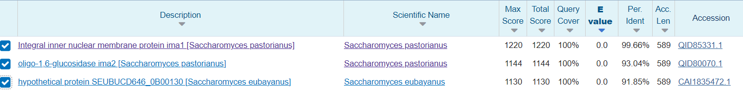
In this step, we are going to take an initial look at differences between the sequences of our query IMA1 protein (S. cerevisiae), and those from S. eubayanus and S. pastorianus.
This takes just two steps:
2. Click on the Alignment tab!
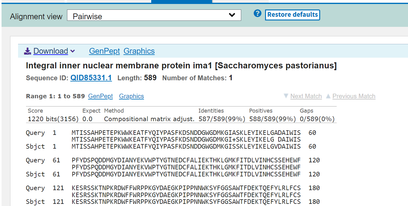
By default, the alignment output is a plain Pairwise alignment. Each section of the page represents one of the three proteins we selected aligned to the S. cerevisiae sequence.
We can “eyeball” some differences, such as the one I highlighted above, but we can make some adjustments to better spot the differences using the Alignment View Dropdown menu:
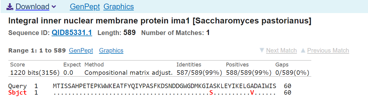
Pairwise with dots for identities: Alignments are anchored (shown in relation to) to the query sequence in pairwise fashion with mismatches colored in red. Sbjct will be in red and bold font if a line in the alignment contains mismatches.
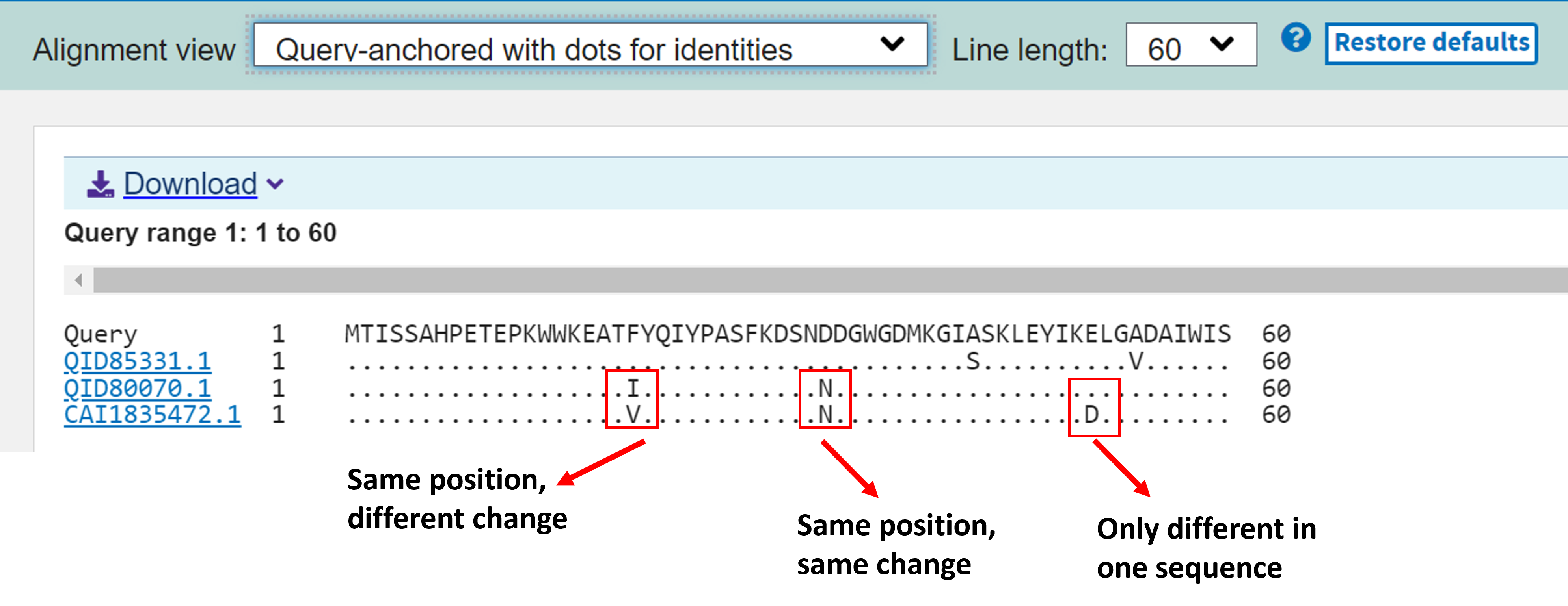
Query-anchored with dots for identities: Displays all result sequences we selected in relation to the query sequence. Identities are displayed as dots (.), with mismatches displayed as single letter abbreviations.
Exploring an Alignment with NCBI MSA Viewer
To enter an interactive MSA viewer with your query and three selected sequences, go back to the "Descriptions" tab, and select the rightmost option: MSA Viewer. You should end up on a screen that looks like this one. A couple of important options are highlighted here: 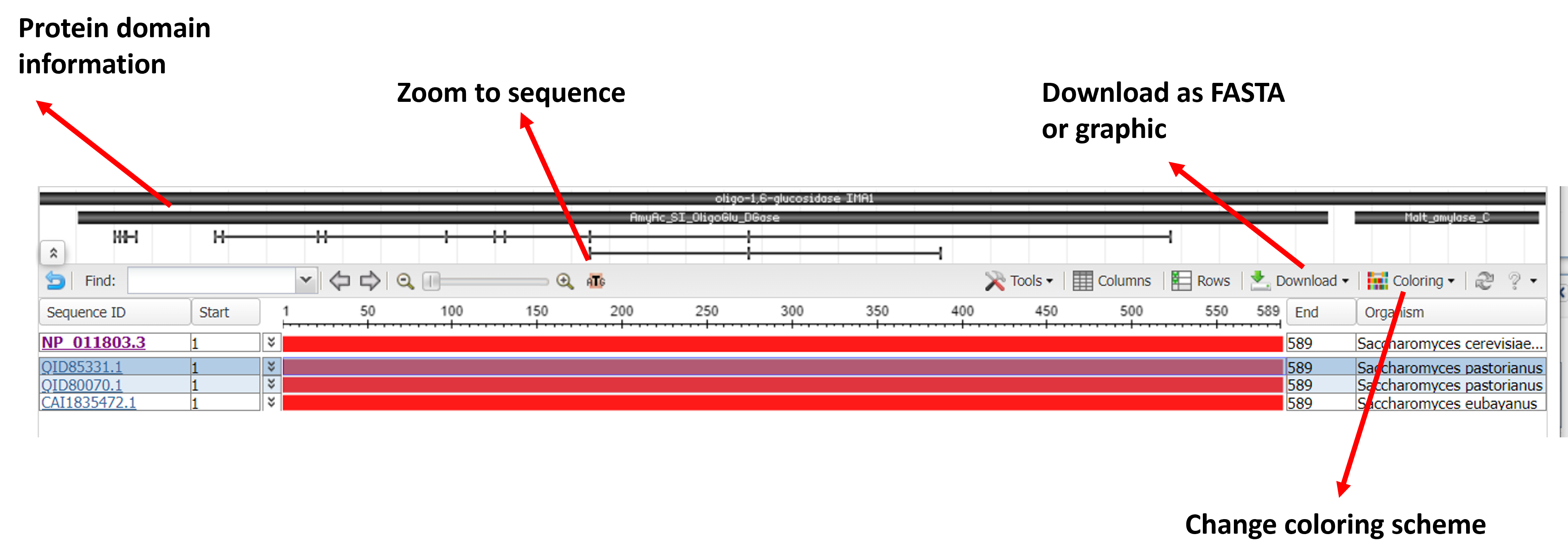
One coloring scheme option best suited for looking at variation is "Color by Differences", which highlights the differences between the subjects and the query sequence. 
You can also zoom in to sequene level to look at individual residues:

Optionally, if you want to download this alignment or a graphic of this alignment - use the Download menu! Or, choose "Share This View" to get a sendable link to this exact view!
![]() What do you notice about the two S. pastorianus sequences? How might these patterns of variation relate to the hybrid origin of this yeast species?
What do you notice about the two S. pastorianus sequences? How might these patterns of variation relate to the hybrid origin of this yeast species?
Finding a 3D Structure for IMA1
First, lets go back to the RefSeq protein page for the original S. cerevisiae IMA1 protein. You may notice a link to a Structure database entry on the righthand side of the page - Click it!
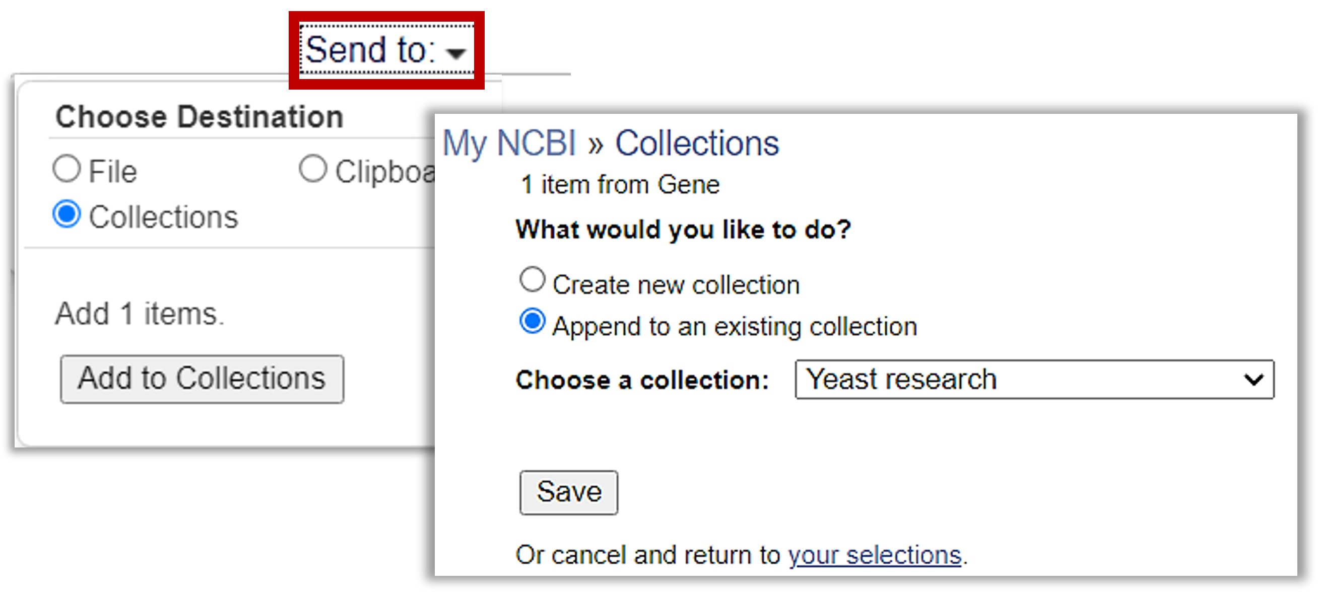
Save the Structure record to the Yeast research folder!
1. Click "Send to" to add to the existing Yeast research folder.
  |
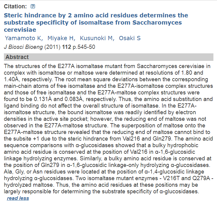
Next, click the associated link to go to into the actual Structure record for the Crystal Structure Of Isomaltase In Complex With Maltose
Once again, this a page with a lotttttt of information. The top part of this page provides some background and information to help you determine if you are on the right page, such as an abstract for the citation related to this MMDB entry.
What did the authors learn about IMA1 in this mutant? Can it bind both maltose AND isomaltose?
 For a hint about what to look for, click here.
![]() For future work, you could click on any number of download links
For future work, you could click on any number of download links
(such as those under Download Structure Data) links
to download the data to a folder on your computer!
The molecular components table is also a rich source of information and links out to other databases, and ways to search for similar domains and molecules:
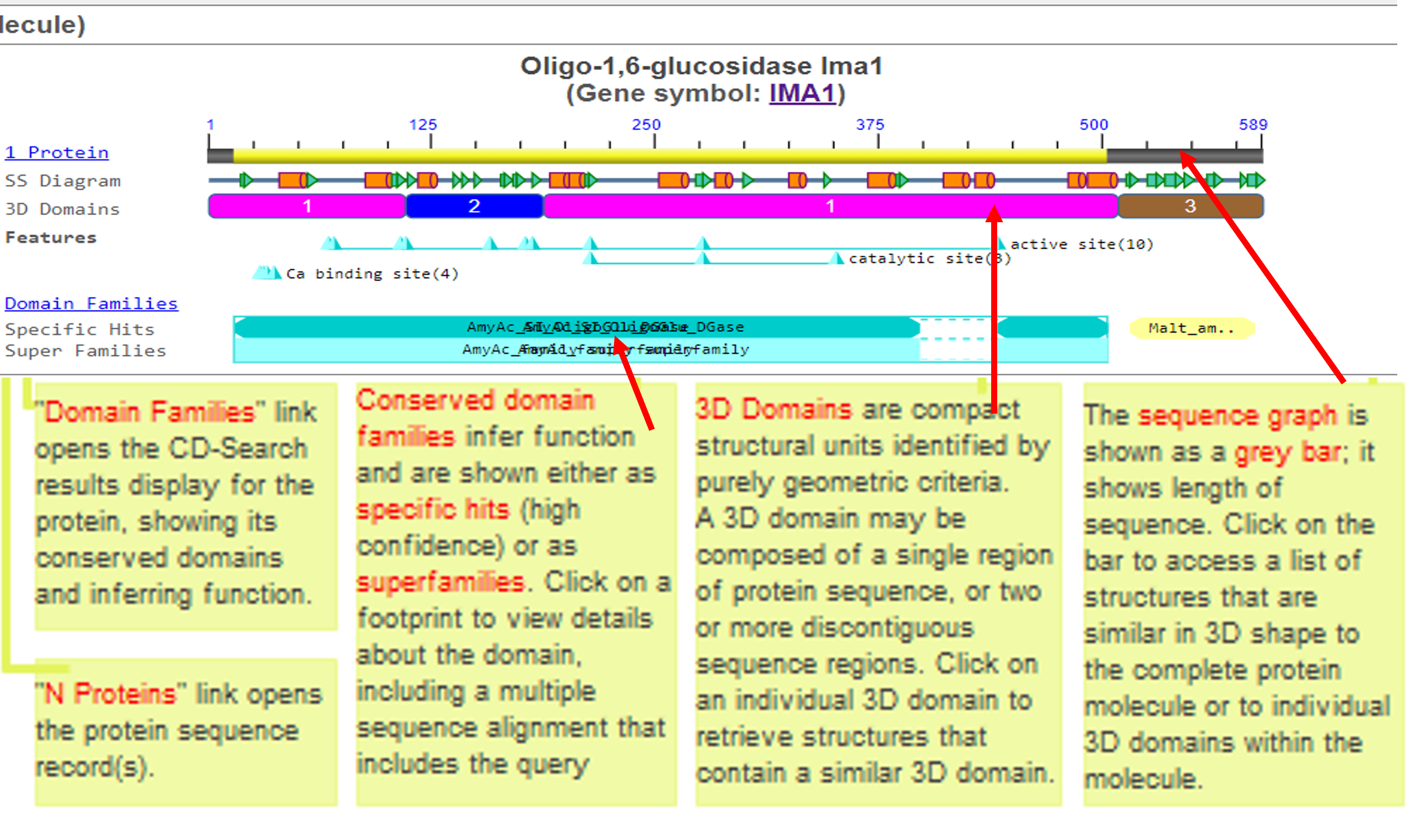
For more information about how to make sense of all the data on this page, MMDB has an extremely thorough of these pages in their help documentation.
Advanced: Interactive 3d Structure with iCn3D
 To see a fully interactive, customizeable version of the chemical structure of IMA1, click on this button!
Advanced Exploration: Align an S. pastorianus sequence to this Structure and visualize
If you want to explore, click this link for a pre-modified 3d structure of IMA1 with this protein aligned and the sugar pocket's side chains rendered and visible!
This is just the very start of what you can do with iCn3D, and there is a lot of help available: |
Last Reviewed: June 29, 2023